Posts

Blood Gas Partition Coefficient

 It is the ratio of the amount of anaesthetic agents present in the blood compared to gas when two phases are of equal volume and pressure and in equilibrium at 37 degrees Celsius.  ⇒ Also called as Ostwald coefficient of blood gas.  ⇒ It expresses the solubility of the agent in blood.  ⇒ The higher the blood/gas partition coefficient, the more the agent is soluble. It will take longer to raise the agent's partial pressure in the blood.  Anaesthesia is related to the partial pressure of the anaesthetic agent at the effect site.  ↑ B/G partition coefficient:  ↑ Duration Onset time  of anaesthesia  ↑ Duration of Offset time of anaesthesia  ↓ B/G partition coefficient:  ↓ Duration of Onset time ↓ Duration of Offset time  The blood gas partition coefficient of the volatile agents: Halothane: 2.4  Isoflurane: 1.4 Sevoflurane: 0.6  Desflurane: 0.4  Halothane is present in the blood 2.4 times more than that present in t...

Clinical effects of Sevoflurane

Due to the preferable pharmacodynamics, Sevoflurane is the most favoured drug for the induction and maintenance of anaesthesia. Its low blood-gas partition coefficient helps to rapid alteration of the level of anaesthesia.  CVS effects:  → Mild ↓ cardiac contractility (dose-dependent, equal to isoflurane) → Mild ↓ SVR (dose-dependent, equal to Halothane) → Mild ↓ BP (dose-dependent, most least compared to others) → Minimal effect on HR → May cause prolongation of the QT interval  → Not associated with coronary steal  → Doesn't sensitize the myocardium  → No effect on splanchnic blood flow  Respiratory effects: → Non-irritant  → Dose-dependent Tidal volume depression (less than Isoflurane, equal to Halothane)  → ↑ RR (equal to Isoflurane, more than Halothane)  → ↓ MV causing ↑ PaCO2 → Depression of hypoxic and hypercapnic ventilatory drive  → Inhibition of hypoxic pulmonary vasoconstriction  → Bronchial smooth muscle r...

Adverse effects of suxamethonium

    Muscle pains  Especially in patient who is ambulant soon after surgery, such as the day-case patient.  Caused by the initial fasciculations, are more common in young, healthy patients with a large muscle mass.  Occur in unusual sites, such as the diaphragm and between the scapulae, and are not relieved easily by conventional analgesics.  The incidence and severity may be reduced by the use of a small dose of a non- depolarising NMBA given immediately before administration of suxamethonium (e.g. atracurium 0.05 mg/kg). However, this technique, termed precurarisation or pretreatment, reduces the potency of suxamethonium, necessitating the administration of a larger dose to produce the same effect.  Increased intraocular pressure   Caused by the initial contraction of the external ocular muscles and internal ocular muscles after administration of suxamethonium.  It is not reduced by precurarisation.  The effect lasts for as ...

Basic physiology of Neuromuscular junction

  Macro-anatomy of neuron ⍺ Motor neuron is surrounded by myelin sheath which is formed by the Swann cell.  Speed of conduction in ⍺ neuron is faster (50 - 100 m/s) due to myelin insulation and node of Ranvier. In the node of Ranvier, there is a high concentration of voltage-gated sodium channel causing depolarisation and saltatory movement to the next node.   The neuromuscular junction commences at the nonmyelinated nerve ending.  Extraocular, laryngeal and some facial muscles are innervated by slow conducting Ɣ neuron with multiple innervations.  Motor endplate  The axon terminal surrounded by Schwann cell cytoplasm contains mitochondria and vesicles.  The synaptic vesicles are synthesised in the anterior horn cell of the spinal cord and transported to the motor nerve terminal via the micro-tubular system.  The synaptic gap is 50 nm wide and contains a basement lamina of 20 nm consisting of mucopolysaccharides.  Ach receptor...

Monitoring neuromuscular block

Image
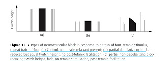
Following stimulation of the relevant nerve. Nerve stimulators must generate a supramaximal stimulus (60–80 mA) to ensure that all the composite nerve fibres are depolarized. Duration 0.1 ms. Negative electrode directly over the nerve. Positive electrode  placed where it cannot affect the muscle in question. Five main patterns of stimulation Single twitch stimulation Simplest form Requires a baseline twitch height Reduction in twitch height observed until 75% of NMJ receptors have been occupied by muscle relaxant  Only a small number of receptors are required to generate a summated mini end-plate potential, which triggers an action potential Partial NMJ block with depolarizing muscle relaxants (DMRs) and non-depolarizing muscle relaxants (NDMRs) reduce the height of single twitch stimulation. Tetanic stimulation Individual stimuli are applied at a frequency >30 Hz Twitches observed in the muscle become fused into a sustained muscle contraction – tetany Most stimulators deli...

Complications of Chronic kidney disease (CKD)

  CNS: Peripheral neuropathy Autonomic neuropathy Encephalopathy  CVS: Fluid overload Hypertension, Left ventricular hypertrophy  Congestive cardiac failure  Uremic pericarditis  Accelerated atherosclerosis with stiffening of vessels  Vascular calcification  Valvular heart disease  Arrhythmia and conduction block  Cardiovascular autonomic neuropathy with  ↓ Baroreceptor sensitivity  Sympathetic nervous system hyperactivity  Parasympathetic dysfunction  Complications due to A - V fistula -  Congestive cardiac failure  Limb ischemia  Steal phenomena  Pulmonary atheroembolism  Pulmonary: Interstitial oedema  Alveolar oedema  Pleural effusion  Hyperventilation  GIT: Anorexia, Nausea, Vomiting  Delayed gastric emptying, Adynamic ileus  Hyperacidity, Mucosal ulceration, Hemorrhage  Metabolic: Metabolic acidosis  Hyperkalemia  Hypermagnesemia  Hyperuricemi...

Causes of Syndrome of inappropriate antidiuretic hormone secretion (SIADH)

 Pulmonary disease  Pneumonia  Tuberculosis  Abscess Malignancy Lung GI tract  Genitourinary  CNS Tumour  Infection  Haemorrhage Drugs

Drugs causing hyponatremia

 ↑ ADH secretion -  Barbiturate  Opioid  Hypoglycemic SSRI Antipsychotic  Carbamazepine  Potentiation of ADH -  Paracetamol NSAIDs ADH analogue -  Oxytocin Vasopressin Desmopressin 

Causes of hyponatremia

  Hypovolemic hyponatremia: Renal loss: Diuretics Ketonuria Hypoadrenalism - mineralocorticoid deficiency  Metabolic alkalosis  Renal tubular acidosis Protein-losing nephropathy  Extrarenal loss: Vomiting  Diarrhoea  Burn  Pancreatitis  Third space loss Hypervolemia hyponatremia: Congestive cardiac failure  Cirrhosis  Nephrotic syndrome  Euvolemic hyponatremia: ↓ Plasma osmolarity: SIADH Renal failure  Hypothyroidism  Surgical stress/ Sympathetic stimulation  Glucocorticoid deficiency Drugs ↑ ADH secretion -  Barbiturate  Opioid  Hypoglycemic SSRI Antipsychotic  Carbamazepine  Potentiation of ADH -  Paracetamol NSAIDs ADH analogue -  Oxytocin Vasopressin Desmopressin  Normal plasma osmolarity: Pseudohyponatremia   Reference:  Smith and Aitkenhead's Textbook of Anaesthesia, 7th Edition Stoelting's Anesthesia and Co-Existing Disease , 7th Edition  Morgan & Mikha...

Coagulation pathway (classical)

Image
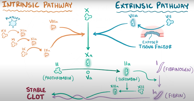
  Extrinsic pathway -   Tissue factor activates FVII. Intrinsic pathway Platelet acts as the stimulating factor, activating FXII, FXI, and FIX. Both intrinsic and Extrinsic pathway follows the common pathway where FX is activated.  Activated FX converts Prothrombin to Thrombin.  Thrombin converts Fibrinogen to Fibrin, which in turn forms the stable clot. Photo credit: Osmosis 

Regulation of Hepatic blood flow

  Blood supply of liver  25% of cardiac output, 1.5 L/ min Blood supply by hepatic artery (1/3 supply, 98% O2 saturation) and hepatic portal vein (2/3 supply, 70 - 85% O2 saturation) The hepatic triad is formed by the branches of the hepatic artery, hepatic portal vein and bile canaliculus which run together  Portal venue and hepatic arteriole join together to form hepatic sinusoid (specialised capillary system) which optimises exchange with hepatocyte  Venous drainage to IVC via right and left hepatic vein  Regulation of hepatic blood flow  Hepatic artery - extrinsic and intrinsic regulation mechanism  Portal vein - extrinsic regulation  mechanism  Intrinsic mechanism:  Myogenic autoregulation 👉🏻 if drop-in hepatic arterial pressure ➡️ flow is maintained by a decrease in hepatic arterial resistance  Hepatic arterial buffer response 👉🏻 if reduction of flow in portal vein ➡️ compensatory decrease in resistance of hepatic artery t...

Anesthetic implications or considerations in Obstructive jaundice

  CVS: Cardiac depression Bile acid causes direct negative inotropic and chronotropic effect SA node inhibition by ↓ spontaneous depolarisation  ↓ Duration of the action potential by inhibiting calcium current  The negative chronotropic impact is due to the binding of Bile acid with the Muscarinic M2 receptor that inhibits intracellular cAMP. ↑ Vagal response  ↑ Plasma ANP levels that inhibit sympathetic transmission and augment vagal response Intravascular volume depletion Due to the effect of Bile acid with Na/H antiport, ↓ Sodium absorption in proximal convoluted tubule and ↑ excretion is accompanied by ↓ water reabsorption.  ↑ ANP and BNP  - diuretic and natriuretic effect  Vascular hyporesponsiveness Endotoxemia induced ↑ vascular Nitric oxide synthesis  Bile acid-induced vascular potassium channel activation  Downregulation of ⍺1 receptor Impaired Baroreceptor sensitivity  Renal: High of acute renal failure due to -  Endotoxem...

Liver or Hepatic Anatomy and Physiology for anesthesiology

The anesthesiologist's most critical aspect of liver anatomy is its blood supply.  The liver derives its blood supply from the hepatic artery and portal vein. These two blood vessels receive about 20%–25% (≈1500 mL/min) of cardiac output.  The hepatic artery provides approximately 25% of the blood flow to the liver, with the portal vein providing the remaining 75%.  The portal vein receives blood specifically from the stomach , intestines , pancreas, and spleen and carries it into the liver through the porta hepatis. While there may be some variations between individuals, the hepatic portal vein is usually formed by the convergence of the superior mesenteric vein and the splenic vein , referred to as the splenic-mesenteric confluence. The hepatic artery arises from the common hepatic artery, a branch of the celiac artery. It runs alongside the portal vein and the common bile duct to form the portal triad.  Owing to the difference in oxygen content of portal venous ...

Drugs associated with hepatitis and hepatic dysfunction

  Toxic effect Alcohol Acetaminophen  Salicylate  Tetracycline  Idiosyncratic effect Volatile anaesthetic (Halothane) Indomethacin  Phenytoin  Rifampicin Toxic + Idiosyncratic effect Amiodarone  Sodium valproate  Methyldopa  Isoniazid Cholestatic effect Oral contraceptive  Anabolic steroid